Quality Control
Find folds, blur, misregistered cycles and segmentation errors, mark them as regions, and flag the cells they affect.
Quality Control marks the parts of an image you should not trust (a fold, a blurred field, a cycle that shifted, a mask that merged two nuclei) and flags the cells they affect. Nothing is deleted: each artifact is a region you can see and edit, and each cell gets a call with its reasons.
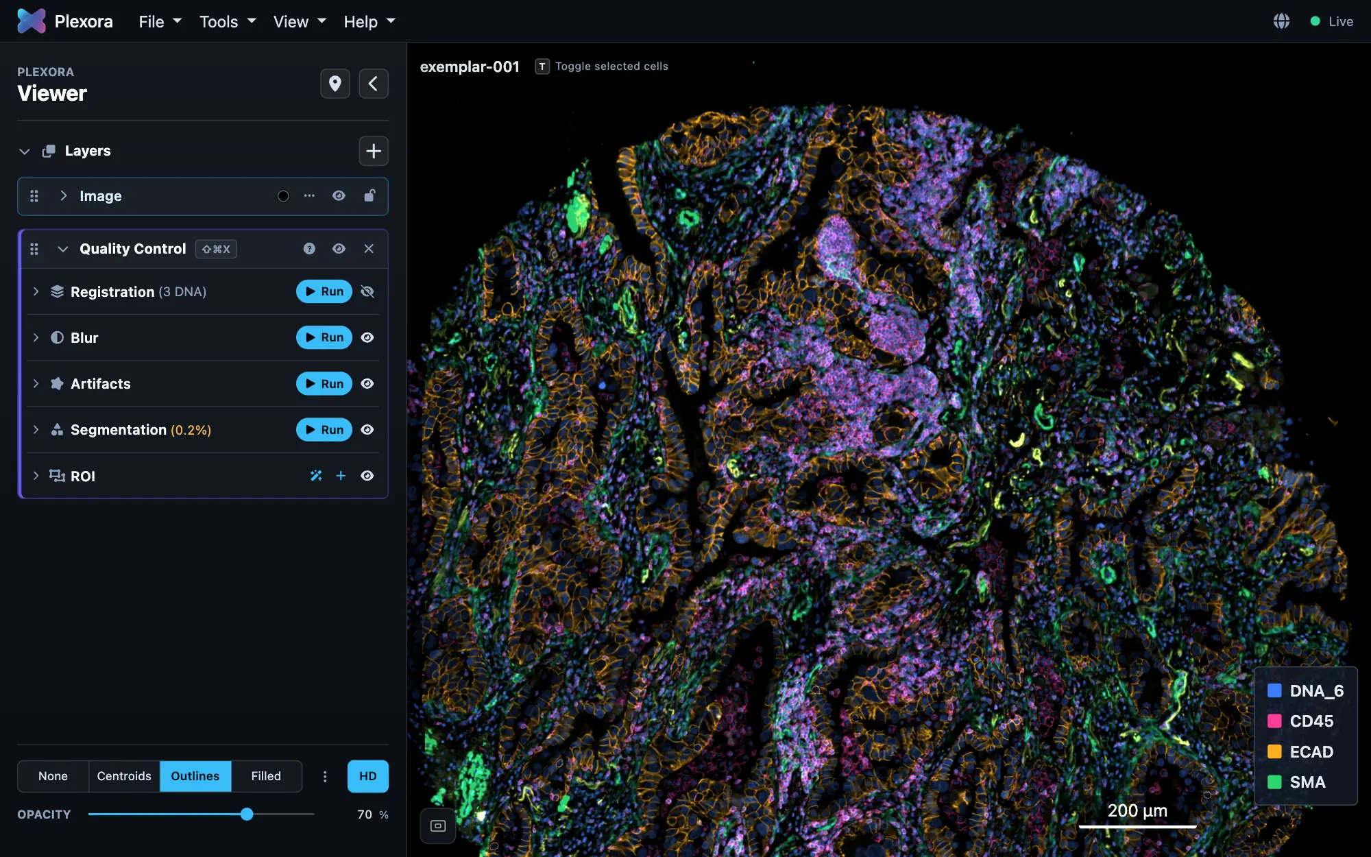
Open it: Tools › Quality Control, or press ⌘⇧X (Ctrl+Shift+X on Windows and Linux). It needs only the image; a cell table adds the per-cell calls.
The card holds five checks, each folded under its own header with a Run button and an eye: Registration, Blur, Artifacts, Segmentation and ROI.
How to use it
Mark a region by hand
In ROI, press Add ROIs and pick a category, or type your own name under Custom and press Enter. The drawing bar appears on the image, the same one the ROI panel uses: draw round the artifact with Polygon, Freehand or Rectangle, or pick magic select (the wand, or E) and drag a Box round it. Click the category on the bar to mark something else, and × when you are done.
Run the automatic checks
Press Run on Blur (out-of-focus tissue), Artifacts (folds, tears, debris, saturation), Registration or Segmentation. Each draws what it found on the image and lists it under its header.
Choose how strict
Lenient excludes only severe, confident artifacts, Standard moderate and severe ones, Strict every confirmed artifact. Changing it recomputes every cell's call; regions you drew keep what you chose.
Review
Click a region to fit the view to it. A region's ⋯ menu has Details (why it was flagged), Trace outline and Delete region…. Click a row under a category to show the cells it flagged.
The checks
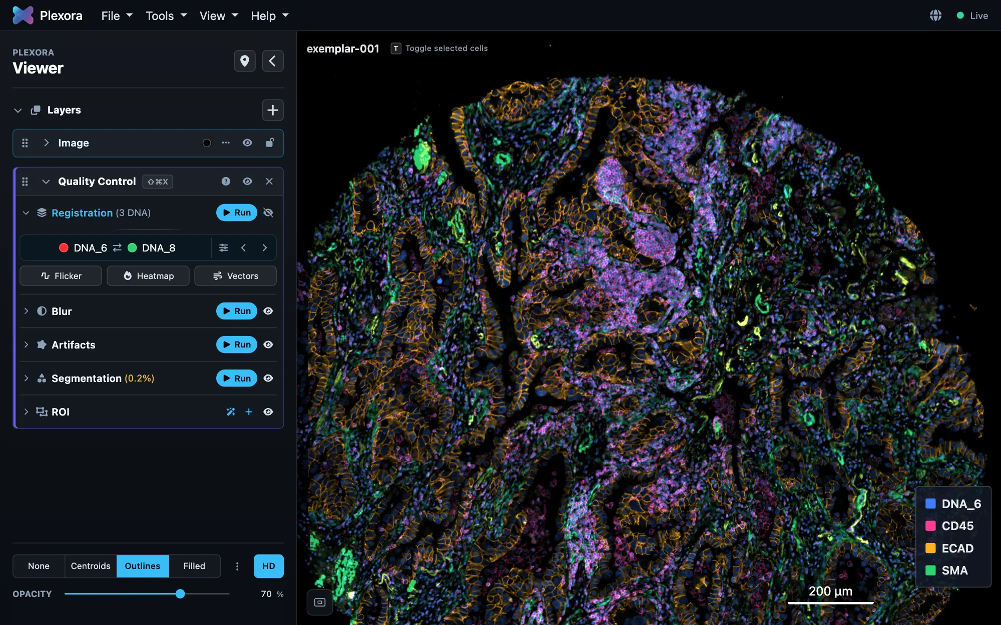
- Registration compares each nuclear channel (DAPI, DNA, Hoechst) with the reference cycle. The channel being compared sits in channel 2, the reference in channel 1; stripes crawl over areas displaced past the threshold. F turns the stripes on and off, Z / X step the comparison channel. The foot of the card shows the open check's keys as buttons: these three for Registration, E for ROI. A heatmap and an arrow field show the shift across the tissue.
- Blur scores the tissue from sharp to blurred against the image's own sharpest tissue, as a heatmap.
- Artifacts finds folds, tears, debris and saturation, one threshold slider per category.
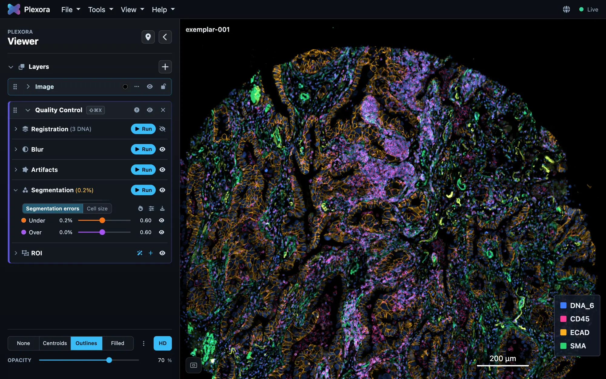
- Segmentation (needs a mask) compares the mask with the DNA stain: Under is one label holding two nuclei, Over is one nucleus cut across two labels. The density map adds Large, Small and Irregular cells.
Tips
- QC regions are ordinary ROIs. Open the ROI tool to rename, reshape or recategorise them; QC takes the edits in.
- A filled dot excludes, a ring only flags. Eyes hide what is drawn, never what is counted.
- Thresholding's Auto threshold leaves out cells QC excluded.
What it saves
QC results are saved with the project. The download button in the card's header offers:
- Cells (CSV): every cell's status, a column per category, and its reasons. Cells (AnnData, obs only): the same as an
.h5adto join onto your own. - Regions (GeoJSON), Provenance (JSON) and Findings (CSV): every region and reason, with its score and threshold.
- Save into file…: adds QC's columns to the table this project was opened from, after asking, with a backup.