Image viewer (2-D and 3-D)
Pan and zoom a whole slide, rotate and flip it, save views, walk a dataset, and look through a z-stack in 2-D or 3-D.
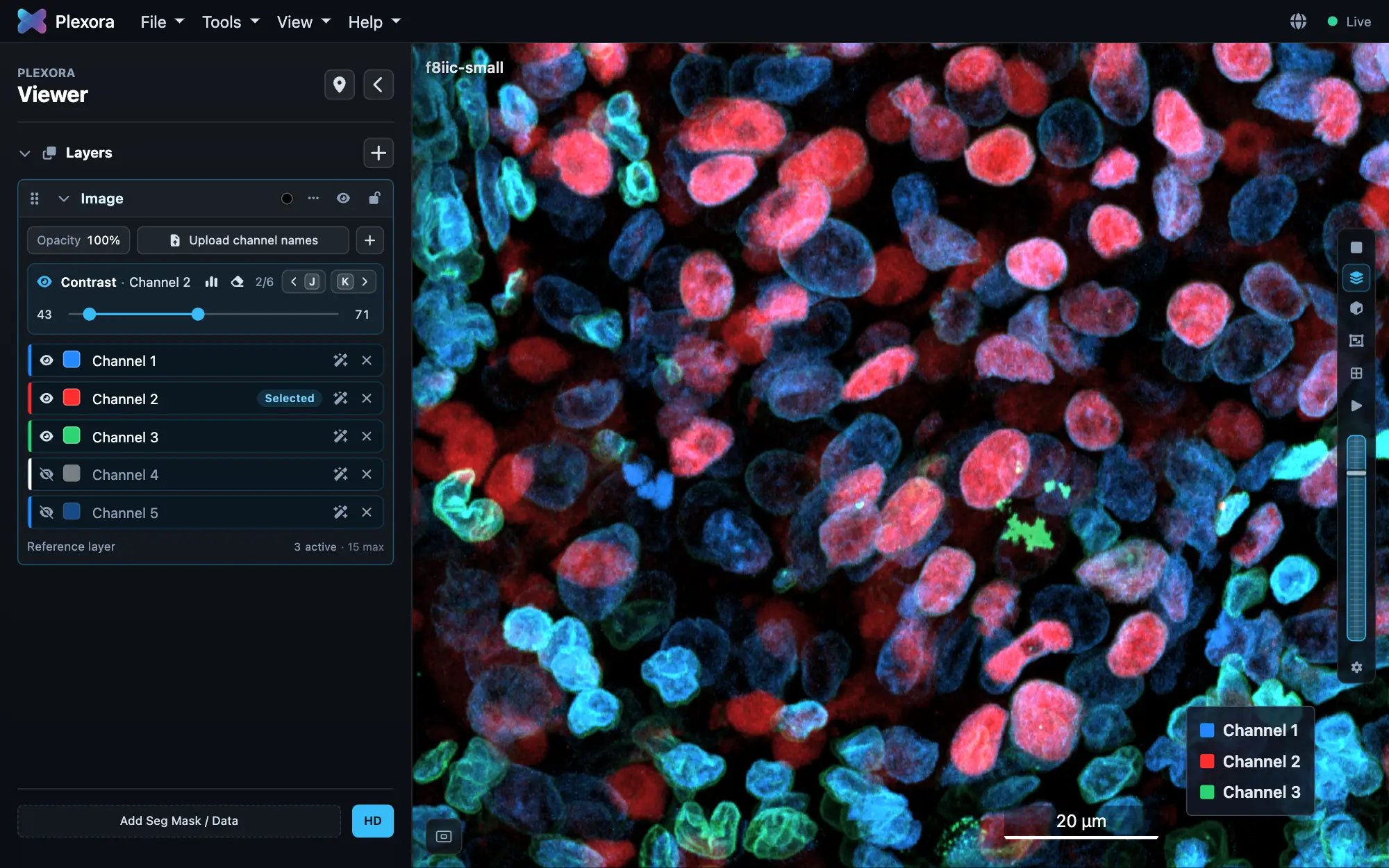
The viewer draws the whole image at every zoom level. A z-stack or a time series adds a depth strip along the bottom, where you switch between one plane, a projection and the stack in 3-D.
How to use it
Move around
Drag to pan and scroll to zoom. The buttons on the left edge zoom and return Home (the whole image). The round button in the bottom-left corner opens an overview map of the tissue.
Turn the view
View › Rotate & Flip turns the image by 90° steps or any angle and mirrors it. The orientation is saved with the sample. View › Scalebar shows or hides the scale bar.
Save a view to come back to
Press M to save what is on screen: position, zoom, channels and contrast. Saved views in the sidebar header opens the tray; click a view to return to it. Saved views are kept by Figure Builder, so they can also go into a figure.
Look through a z-stack
On the depth strip, choose One plane, Projection of a range of planes or The stack in 3-D (V). Step planes with ] and [ (with Shift, ten at a time), and frames with . and ,. Sections adds XZ and YZ views beside the image.
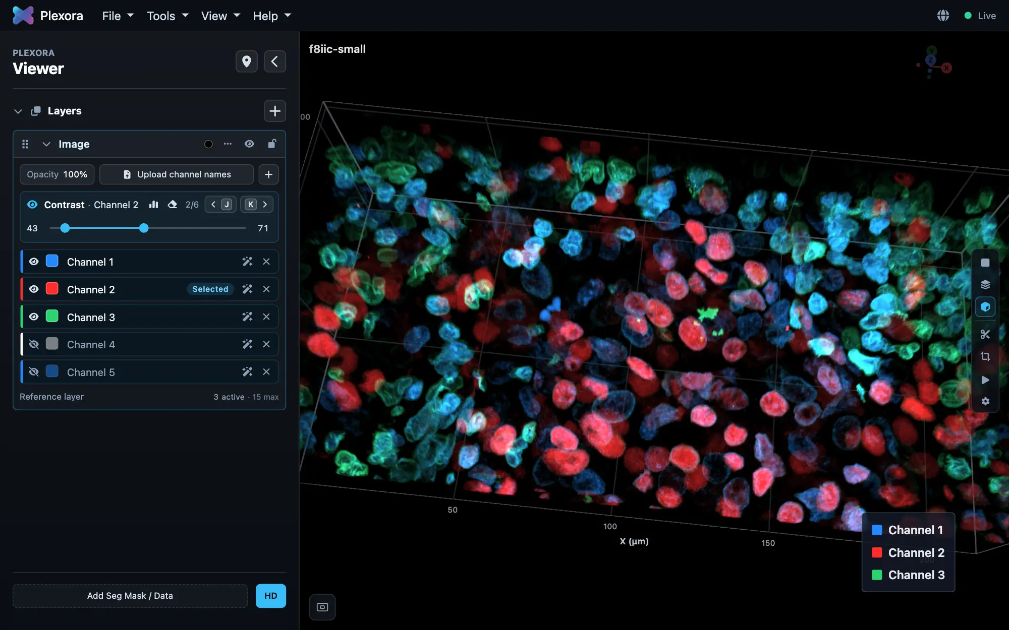
Open up the stack in 3-D
Drag to turn the stack. Cut takes a side off it and Crop keeps a box of it (O shows or hides the cut panel). Area places a square on a plane and opens just that part in 3-D.
Tips
- HD, in the sidebar's Cells row, reloads the image at full 16-bit precision for judging faint signal. Leave it off for ordinary browsing.
- In a dataset, Previous / Next at the top right (or B / N) opens the neighbouring sample and carries over your channels, colours, cell display and open tools. Contrast and gates stay each sample's own.
- In 3-D, X, Y or Z limits turning to that axis, and 0 returns to the angled default view.
- A brightfield slide has Brightness, Contrast and Gamma sliders on its Image card instead of channels. They change the display only and are not saved.